Snake Bites – First aid, Treatment and Symptoms
Mar 27, 2026
Last Updated On: February 9, 2026
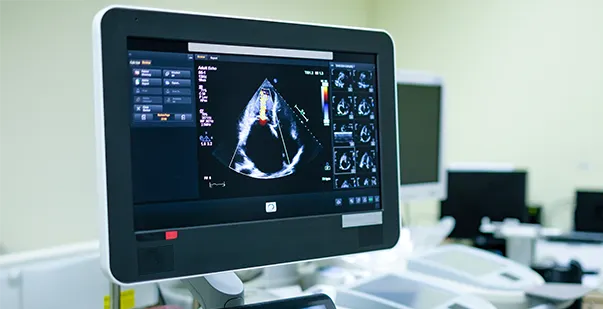
Your heart beats about 100,000 times a day, pumping blood through a vast network of 60,000 miles of blood vessels. But how do you know if it’s working properly? That’s where an echocardiogram comes in. This non-invasive test uses sound waves to create detailed images of your heart in real-time, which helps doctors assess its structure, function, and blood flow.
Heart disease remains the leading cause of death worldwide, yet many people don’t realize they have a heart problem until symptoms appear. An echocardiogram can detect issues like weak heart muscles, valve disorders, or abnormal blood flow before they become serious.
Over 7 million echocardiograms are performed in the United States annually to manage heart diseases and prevent complications. Now, let’s understand more about these tests and their role in ensuring improved heart health.
An echocardiogram is an ultrasound test to check the structure and function of your heart. It can diagnose multiple health conditions, including cardiomyopathy and valve diseases. During an echo test, your healthcare provider uses ultrasound from a hand-held wand placed on your chest. It takes pictures of your heart’s valves and chambers immediately. This cardiovascular assessment helps the provider analyze the pumping action of your heart.
They may also give you some effective tips to avoid a heart attack and improve your health after the echo test. A standard echocardiogram is a painless, simple, and safe procedure. There are no side effects from the scan, but the lubricating gel may sometimes feel cold. You may also experience some minor discomfort when the electrodes are removed from your skin at the end of the echo test.
Unlike X-rays, CT scans, and other diagnostic tests, an echocardiogram does not involve any radiation. This common test can easily show blood flow through your heart and the adjacent valves.
A technician called a cardiac sonographer usually performs your echo test. These professionals are trained in performing echocardiograms and use the most current technologies.
Healthcare professionals use different types of echocardiograms to conduct a heart disease diagnosis on patients. The type of test you need for the analysis of your heart functions depends on the choice and requirements of your healthcare provider.
The common types of echocardiograms are:
The TTE is a standard echocardiogram and is also called the heart ultrasound. It’s a noninvasive test that analyzes blood flow through your heart and the heart valves. A TTE assists healthcare professionals by providing accurate pictures of the heart from outside the body. It allows your heart’s structures to show up better on the images for doctors to diagnose all underlying issues.
You may need to undergo a TEE test if a standard echocardiogram doesn’t provide relevant details about your heart valve functions. It gives a detailed analysis of the heart and the body’s main artery, called the aorta. A TEE also captures images of the heart from inside the body. Please note that this test shouldn’t be done if you have bleeding in the upper gastrointestinal tract or a tear or tumor in the esophagus.
This echocardiogram test is done during pregnancy to check the baby’s heart. It’s a noninvasive test that involves moving the ultrasound wand over the pregnant mother’s belly. This enables the healthcare professional to see the unborn baby’s heart with cardiac imaging, which is a series of techniques to diagnose cardiovascular conditions.
A stress echocardiogram is done before and after you exercise in any hospital setting. It analyzes how your heart responds to physical activity or stress. A healthcare professional may order this test if they think you may have coronary artery disease. You may also be given medicine that affects the heart like exercise does if you cannot indulge in any physical activities.
Healthcare professionals leverage echocardiograms to see the structure, size, and activity of your heart. It helps them diagnose heart problems and decide on their next actions to manage their health. More specifically, professionals use echocardiograms to diagnose the following:
Proper preparation ensures accurate results and a smooth experience during an echocardiogram. While most cardiovascular assessments require minimal preparation, specific types, such as stress echocardiogram or transesophageal echocardiogram (TEE), may have additional guidelines. Following these steps can improve the test’s effectiveness.
Your healthcare provider may provide specific guidelines based on your medical history and the type of echocardiogram. Always confirm any special requirements before the test.
If you are scheduled for a transesophageal echocardiogram or a stress echocardiogram, you may need to avoid food and drinks for several hours before the procedure. This helps ensure clear cardiac imaging and accurate readings.
Inform your doctor about any medications you take, especially blood pressure or heart-related drugs. Some medications may need to be temporarily stopped or adjusted before the test.
For a standard transthoracic echocardiogram (TTE) or a stress test, wear loose, comfortable clothing. Avoid tight garments and jewelry that might restrict movement. If undergoing a stress echocardiogram, wear comfortable shoes for treadmill exercise.
Certain medications can affect the results. Ask your doctor whether to continue or stop blood pressure, heart rate, or diabetes medications before the test. Never stop any prescribed medication without approval.
If undergoing a stress echocardiogram, avoid caffeine and nicotine for 24 hours before the test. These substances can alter heart rate and affect test accuracy. Decaffeinated drinks may still contain caffeine, so check labels carefully.
An echocardiogram is a non-invasive and painless test, but stress and anxiety can affect heart rate and readings. Stay calm and breathe normally during the procedure for the most accurate results. If you are feeling anxious, talk to your doctor about what to expect. Knowing the steps in advance makes the process smoother.
An echocardiogram provides detailed images and measurements of your heart’s structure and function. You must understand the key aspects of your results to interpret what they mean for your heart health. These include:
The top of the echo report usually includes specific patient details, such as name, age, gender, and test date. Healthcare providers or professionals conducting the test must ensure these reports are accurate and do not lack any information.
The report will also include several images or “views” of the heart and its adjacent valves. These views may consist of parasternal, apical, subcostal, and parasternal windows. Each window view provides a different perspective of the heart and its chambers, blood flow, and valves.
The EF is a key measure of the heart’s pumping efficiency which is expressed in percentage. It usually indicates how well your heart can pump blood. Usually, an ejection fraction falls between 50% and 70%.
The echo test analyzes the heart valve functions, comprising the mitral, aortic, tricuspid, and pulmonary valves. The report also notes whether the valves of your heart are functioning normally or if there is leakage or narrowing that requires immediate medical attention like cardiac massage or other procedures.
The measurements of the heart’s chambers are recorded in an echocardiogram. This involves capturing images of the left ventricle, right ventricle, left atrium, and right atrium. Enlargement of these chambers often indicates certain heart conditions in patients.
The echocardiogram also indicates if there are any areas of your heart with abnormal wall motion. This could suggest heart muscle damage or ischemia, which implies a lack of blood flow.
The cavity that encircles your heart is called the pericardium. If there are any accumulations of fluid around the heart), which might be an indication of inflammation or other diseases, the echo test might highlight them.
Echocardiograms help in the diagnosis, treatment, and management of cardiovascular diseases, which remain a leading cause of mortality worldwide. These tests also assist in preventative cardiac care by enabling healthcare professionals to analyze underlying health conditions accurately.
The following points further highlight the role of these echo tests in preventative cardiac care.
Cardiac imaging techniques are an inevitable part of echocardiograms. They enable healthcare providers to detect and diagnose heart conditions at an early stage when treatment options are the most effective. An echo test helps doctors make timely and accurate diagnoses by identifying structural abnormalities and analyzing heart functions. It also leads to better patient outcomes.
Once a heart condition is diagnosed, an echocardiogram provides valuable information to guide treatment decisions and monitor disease progression. Any healthcare professional can easily determine the most appropriate medication therapies, lifestyle modifications, or surgical procedures for each patient’s unique needs. Electrocardiograms are also useful in evaluating the effectiveness of these medical treatments and detecting potential complications.
Echo tests can assist healthcare providers in analyzing a patient’s cardiovascular risk profile. It also helps them devise preventive strategies to reduce the risk of future cardiac events. Healthcare professionals can easily implement lifestyle modifications, initiate appropriate therapies, and provide patient education by identifying individuals at high risk for heart complications. This also empowers you to take control of your heart health and minimize any risk of adverse health outcomes.
Early detection of heart complications can help you save the lives of several people. The best way to do this is to perform an echocardiogram test on the intended patient. This provides the correct diagnosis of the patient’s heart functions so that they get the perfect treatment plan curated to their requirements. If you’re in the healthcare field, consider undergoing an online cardiopulmonary resuscitation (CPR) course or first aid certification to learn more about such echo tests. This will enable you to interpret echocardiogram results more effectively and respond to cardiac emergencies with confidence.
Read More:
Cardiac Arrest: Who is More Likely to Survive Men or Women?
Subscribe
Search Here
Select Courses
Recent Posts
Snake Bites – First aid, Treatment and Symptoms
Categories